Bacteria and cancer – The diagnostic value of cancer’s microbiome
It has been widely accepted that microbes play essential roles in multicellular organisms, from digestion to immunity and overall homeostasis. With the development of meta-omics, scientists are now able to peek into the diversity of microbial life inside multicellular organisms. Although scientists have just had a glimpse of complex interactions of microbes and their hosts, it is already known that changes in the composition of microbial communities (microbiome) could point to certain diseases.
The microbiome and changes to it have already been used as markers for different diseases, such as inflammatory bowel disease, obesity and cancer.
Read more about how different microbiomes are involved in tumour development, tumour promotion and tumour progression.
In this article we will explore recent insights on how the microbiome can be used for cancer diagnostics and monitoring cancer progress (Messaritakis et al., 2020).
The link between bacteria and cancer diversity
Each tumour type is unique due to different genetic and epigenetic alterations that are mainly induced by changes in the intracellular and extracellular environment.
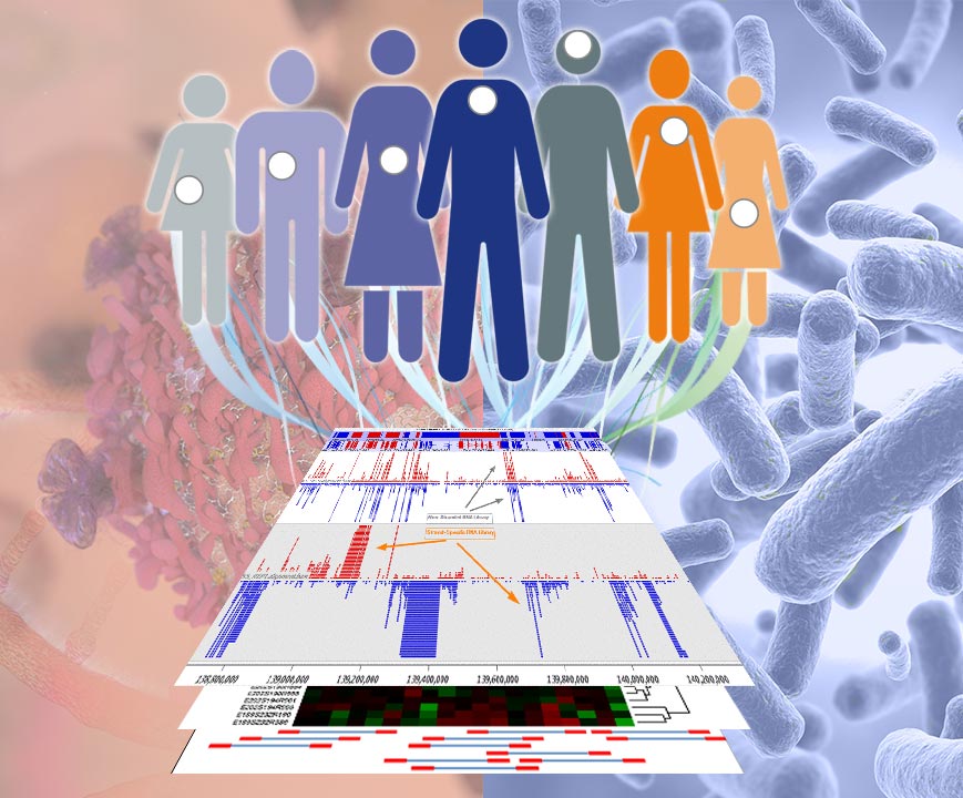
Recently, the Cancer Genome Atlas (TCGA), a database of human cancer mutations, has been published. This ambitious project was initiated in 2005 and aimed to sequence all tumour genomes and their epigenetic states, as well as their transcriptomes and proteomes. Besides cataloguing eukaryotic sequences, many prokaryotic sequences have also been included in the database! Although it has been known for a century that microbes and cancer are linked, this result has spurred a new initiative to determine the atlas of microbes for different types of cancer: The Cancer Microbiome Atlas (TCMA) (Dohlman et al., 2021).
It has been found that the microbiome of certain types of cancers is in some cases very similar to the microbiome of the healthy organ in which the tumour has formed, whereas in other cases, the microbial composition radically differs from the healthy organ tissue. The microbial load (amount of microbes), on the other hand, seems to constantly change throughout tumour progression, especially during late stage cancer.
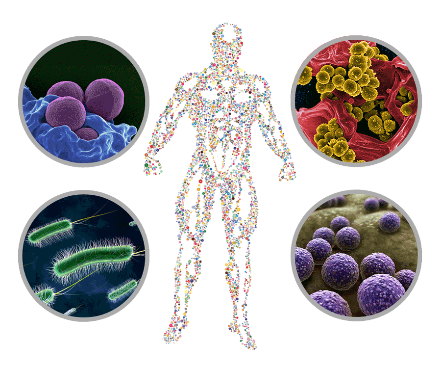
Interestingly, it has been found that most of the present bacteria reside inside tumour cells. As each cancer and its intracellular and extracellular environment is different, it represents a suitable environment for different bacteria. For example, bone malignancies that are characterised by elevated levels of hydroxyproline (amino acid found in collagen) are enriched with bacterial species that can degrade hydroxyproline. Lung tumours are enriched with bacteria that can degrade chemicals found in cigarette smoke, such as toluene, acrylonitrile and aminobenzoates. Alongside local changes to the microbiome, cancer patients usually develop a dysbiosis (imbalance of the microbiome) elsewhere in the body, especially in the gut. The link between cancer progression and gut dysbiosis, however, remains elusive.
Bacteria and cancer: Interdependency of dysbiosis and cancer
In the context of cancer, dysbiosis can either occur in response to cancer or induce carcinogenesis.
In the case of dysbiosis occurring in response to cancer, changes in the microbial composition could be based on the different environmental conditions in a tumour, such as higher amounts of hydroxyproline, acidification or hypoxia.
When it comes to inducing carcinogenesis, bacterial pathogens such as Helicobacter pylori, which can induce peptic ulcer disease (gastric ulcer), have been found.
Interestingly, microbes also seem to be able to alter the progression of cancer by educating the systemic and local immune system. Tzeng and colleagues have demonstrated that several bacterial taxa strongly correlate with the expression of different immunomodulatory genes and immune cell infiltration, for instance. The loss of some of these beneficial bacterial taxa can even cause cancer-promoting inflammation and generally promote carcinogenesis.
Based on the observed differences in the bacterial community of cancer and healthy tissue, researchers investigated the possibility to use bacteria as diagnostic and prognostic markers for cancer (Nejman et al., 2020; Tzeng et al., 2021; Dohlman et al., 2021).
Bacteria and cancer: The prognostic power
Before the microbiome of cancer can be utilised as a marker, the issue of sampling arises.
Sampling of tumour tissue (and residing bacteria) often involves invasive procedures to obtain tissue biopsies. However, there are also minimal invasive or non-invasive sampling methods to determine the bacterial diversity of cancer patients.
The bacteria of patients with colorectal cancer (CRC) have been investigated by blood biopsies. Here, the phenomenon of “microbial translocation”, in which live bacteria cross the mucosal barrier of the intestines and enter the blood stream, is utilised. The extracted bacterial genomic DNA from the blood was used in conventional end-point PCR with primers targeting bacteria specific sites. The results showed significantly higher amounts of bacteria-specific PCR amplicons in CRC patients.
Bacteria-derived extracellular vesicles
Furthermore, bacterial nucleic acids have been found in the blood of healthy people as well as in cancer patients, encapsulated in bacteria-derived extracellular vesicles (EV). These EVs can be used to determine the microbiome composition or abundance of specific bacterial taxa inside the body. Yang et al. created a diagnostic method for diagnosing and monitoring brain cancer based on EVs. The brain is virtually a sterile organ, as the blood-brain barrier prevents microorganisms from entering the brain. Nevertheless, brain cancer patients often develop gut dysbiosis that can be detected by monitoring the change in the bacterial EV secretion. The team’s method relies on extracting bacterial DNA from EVs in the serum of brain cancer patients. Subsequently, the DNA is amplified by PCR targeting the 16S rRNA gene and the resulting amplicons are sequenced, using high-throughput sequencing. The researchers found that in brain cancer patients, in contrast to healthy patients, Dialister and Eubacterium rectale EVs are significantly reduced, whereas Lanchospiraceae EVs are found in much higher levels in the blood. Even though these results do not provide information on the process of EV formation (EVs are either secreted by live bacteria or formed in the process of bacterial cell death), this method represents an innovative and minimal invasive diagnostic tool (Jeong et al., 2020; Yang et al., 2020).
The same research group also developed a test for hepatocellular carcinoma, based on the same diagnostic method. They also showed that the composition of the bacterial community in the same type of cancer differs between stages of cancer progress. Consequently, circulating EVs can potentially be used as biomarkers for diseases themselves, as well as indicators of the severity of the disease (Yang et al., 2020).
Similar results were found in pancreatic cancer patients by Jeong and colleagues. They found that the composition of the bacterial community of cancer tissue differs throughout the stages of pancreatic cancer. The bacterial genus of Tepidimonas was generally more abundant then in normal pancreatic tissue. The exact role of Tepidimonas in cancer tissue is not clear, although some interesting experiments on Tepidimonas fonticaldi showed that it can promote invasiveness and migration of pancreatic cancer cells in vitro. In patients with bigger pancreatic tumours, the genera Leuconostoc and Sutterella were decreased. Patients with additional lymph node metastasis showed significantly increased Comamonas and Turicibacter levels. Comamonas is known to affect the metabolism of cancer patients, and Turicibacter has been linked to other cancers. In terms of tumour recurrence, Streptococcus and Akkermansia, both shown to have some anti-tumour properties, were found to be decreased in tumour tissues (Jeong et al., 2020).
Excursion microbiome analysis: To culture or not to culture
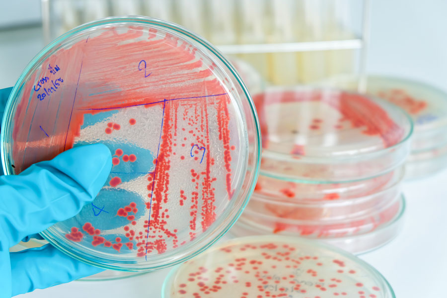
One way to determine the composition of a bacterial community is to culture the bacteria on specific selective media, then isolate and further subculture them. The DNA of the bacterial colonies can then be extracted and analysed. Even though a large variety of growth media that would cater to the specific needs of different bacteria exists, this approach is rather insufficient as most bacteria seem to be non-culturable on these media.
In order to capture as many bacterial taxa as possible, the bacterial DNA is extracted directly from a sample and used in a PCR targeting the 16S rRNA gene. The resulting amplicons are then sequenced via high-throughput sequencing. However, it has been shown that the DNA extraction method and targeted hypervariable region of the 16S rRNA gene both affect the results of the biodiversity profiling (Lim et al., 2018; Teng et al., 2018; Villette et al., 2021).
Bacteria and cancer: Issue of prognostic application and the solution
The biggest problem of utilising bacteria as prognostic and diagnostic markers for cancer is the very low bacterial load in tumours.
In order to solve the problem of very low bacterial load, the detection method needs to be highly sensitive. Methods such as digital PCR (dPCR) and high-throughput sequencing of 16S rDNA amplicons are particularly well suited for these analyses.
Learn more about the application of digital PCR in oncology.
Learn about NGS as clinical diagnostic tool in oncology.
Eventually, changes in the microbiome could function as additional marker for cancer diagnostics. Messaritakis et al. devised a diagnostic method for colorectal cancer (CRC), in which they analysed toll-like receptor/vitamin D receptor (TLR/VDR) gene polymorphisms and detected and analysed bacterial DNA fragments in the blood of CRC patients. TLR and VDR are involved in the detection of microbial antigens and in activating of the immune system against microbes. Polymorphisms in the TLR and VDR genes are linked to carcinogenesis. In this study, significant correlation between the bacterial DNA sequences in the blood of CRC patient and TLR and VDR polymorphisms has been found. This suggests that these polymorphisms have an adverse effect on managing microbial translocation and persistence in the blood, and the involvement of bacteria such as Bacteroides fragilis in cancer development and progression (Messaritakis et al., 2020).
Bacteria and cancer: Conclusion
Changes in the composition of bacterial communities in tissues, also known as dysbiosis, have been found to have great potential as a disease marker, especially in oncology. Here, various types of samples, such as live bacteria or their fragments encapsulated in EVs in blood biopsies, can be used to detect and monitor different types of cancer, even cancers that are difficult to access, such as brain cancer.
Microbiome analysis could be a meaningful addition to already established methods for providing deeper insights and improving diagnostics and monitoring of cancer and its progression.
By Matko Tudor and Dr Andreas Ebertz
Did you like this article about the link between bacteria and cancer? Then subscribe to our Newsletter and we will keep you informed about our next blog posts. Subscribe to the Eurofins Genomics Newsletter.


