
Lion Detect – The self-amplifying disease detection system
“Early detection offers the best chances of cure”. This statement is true for many diseases. Early detection could lead to delaying the onset of the disease and preventing severe progression of the disease, as treatment of the disease can be started earlier and the chances of success are thus greater. However, early detection of diseases is not always possible and is often limited by inadequate diagnostic methods and technologies. Especially during the Corona pandemic, the importance of rapid and easy detection of antigens (in this case the coronaviruses) became clear.
We, the iGEM team Braunschweig, have set ourselves the goal of developing a system that can detect minute amounts of disease-relevant antigens using a self-amplifying system. In addition, a large part of our work is dedicated to raising awareness about oesophageal cancer, as this disease has given us a strong desire to develop such a system. Individuals suffering from this disease show symptoms at a late stage, which in many cases are not associated with oesophageal cancer at first. Our dream is that LionDetect will one day enable rapid and early diagnosis through the detection of small amounts of tumour markers in the blood and thus the treatment of oesophageal cancer.
Due to the relatively small time frame of the iGEM Competition, our project “only” lays the first stones towards this idea.
Our solution and our system for disease diagnostics
We want to design a self-amplifying detection system that can detect the smallest amounts of antigens. For this purpose, our system is divided into three components: The detection component, the reporter component and the amplification component (figure 1). The signal mediated between the three components is a protease activity. This means: if the signal is not present, the protease remains in the two split halves and is therefore inactive. When a signal is detected, the protease is active as the cleaved halves form a complete protease. The active proteases can cut proteins at a specific protease recognition site.
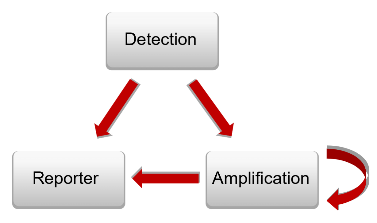
Figure 1: Depiction of the dependence of the individual components of the project with each other
ScFv fragments – The detection component
Here, we work with scFv fragments. ScFv fragments are artificial antibody fragments consisting of antigen-recognising variable domains of the light and heavy chains of an antibody connected by a linker peptide (figure 2). By working with this part in the detection component, any antigen can be detected by replacing the scFv fragment. This makes the application of the system so universal.
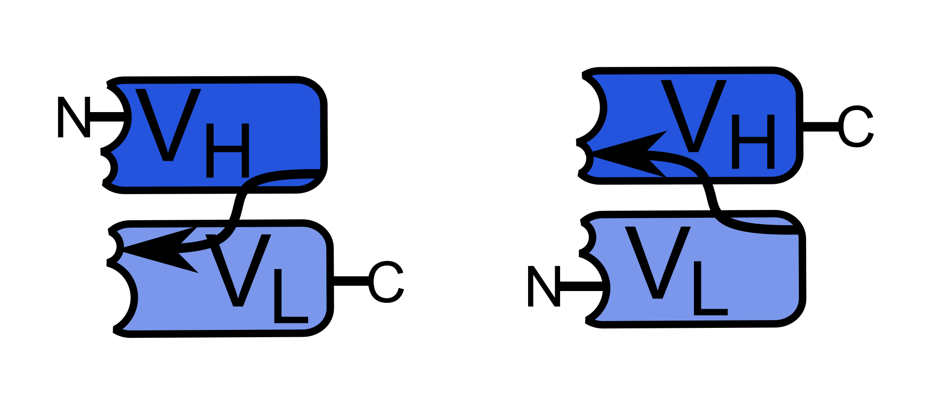
Figure 2: Structure of a scFv fragment, VH represents the heavy chain variable domain, VL represents the light chain variable domain, antigen binding sites are represented by N-and C-termini, arrows represent linker.
The scFv fragments are each bound to a part of a split protease. By binding of the scFv fragment to the desired antigen, the two halves come into spatial proximity so that an active protease can be produced (figure 3).
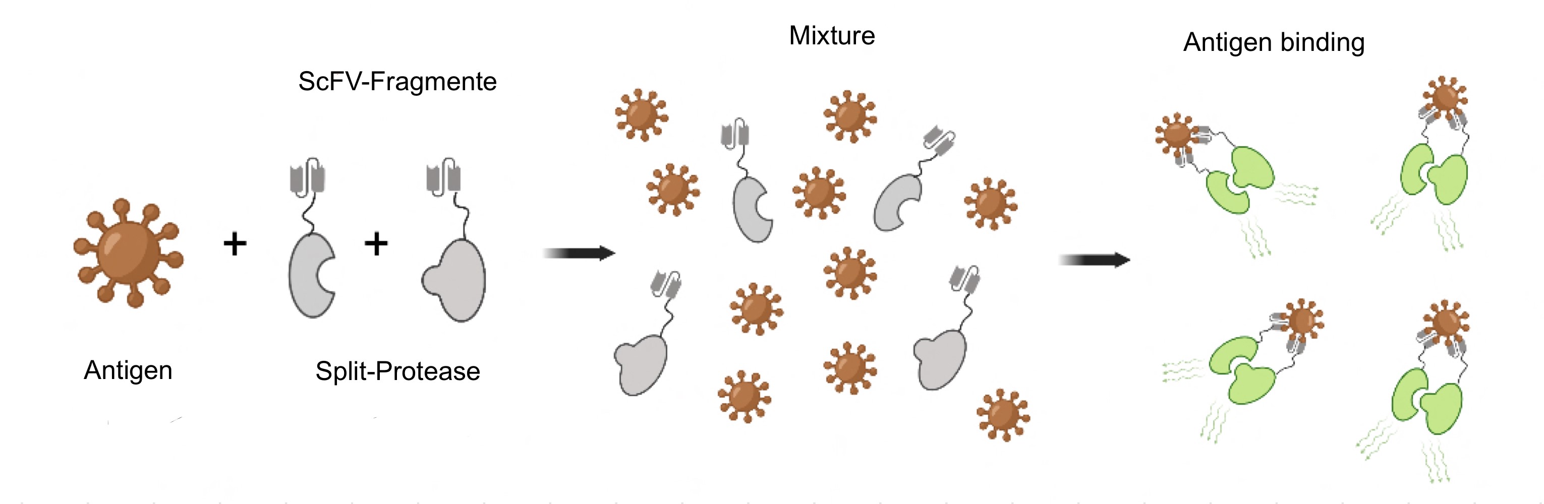
Figure 3: Functionality of the detection component.
ScFv fragments – The amplification component
The amplification is based on a modified intein system. An intein is an amino acid sequence of a protein that can cut itself out of the protein and re-link the remaining parts (exteins) through a peptide bond.
In our project we work with a split variant of an intein. This means that when the C- and N-part of this split intein interact with each other, the intein cuts itself out and the extein part (in our case split-protease halves) are joined together to form an active protease. To ensure that this interaction does not occur without prior activation, the sites of the intein halves that need to interact are masked. We make this possible by attaching a slightly modified part of the C-part of the intein to the N-part of the intein using a linker. The same principle applies to the C-terminal part of the intein and so, this created “cage” can prevent the interaction. By a cut at the protease cutting site in the linker by an active protease (e.g. from the detection component or the amplification system itself), this cage can be detached from the split-intein halves and an interaction between N- and C-terminal intein is ensured. The cutting reaction takes place and an active protease is formed.
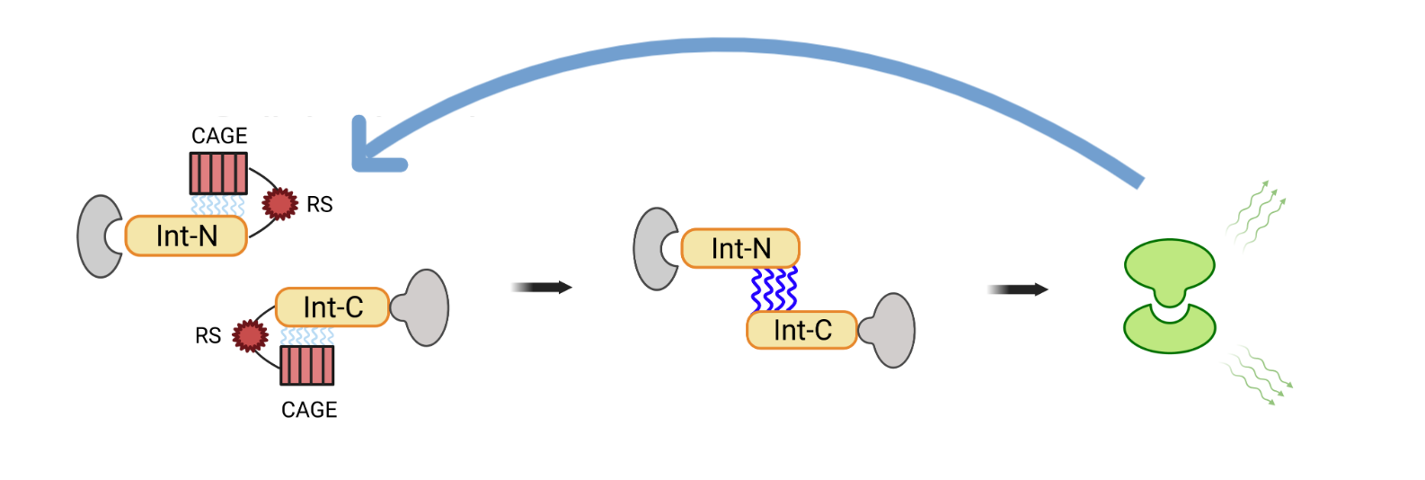
Figure 4: Functionality of the amplification component; Split intein halves are activated by a cut of an active protase in the linker and thus form an active protase; RS= recognition site of the protease.
ScFv fragments – The reporter component
For the reporter component, we work with a fluorescence resonance energy transfer (FRET) pair. This pair consists of two fluorochromes (donor and acceptor; figure 5). Between these, there is a linker with a protease cut site.
By shining light of a certain wavelength on this pair, the emission of the donor can be detected, since the energy gets transferred from the acceptor to the donor. The active proteases of the detection and amplification components can now cut the bond between these two fluorochromes. As a result, the space between the donor and acceptor is too big for energy transfer to take place and the emission of the donor can be detected.
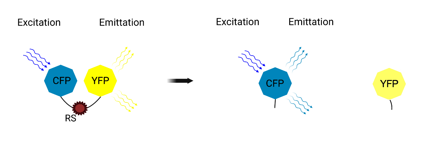
Figure 5: Functionality of the reporter component.
Our aim is that the cell lysates of each of the three components can be brought together so that the desired antigen can be recognised by a detectable colour change.
In the end, our system should work in a cell lysate.
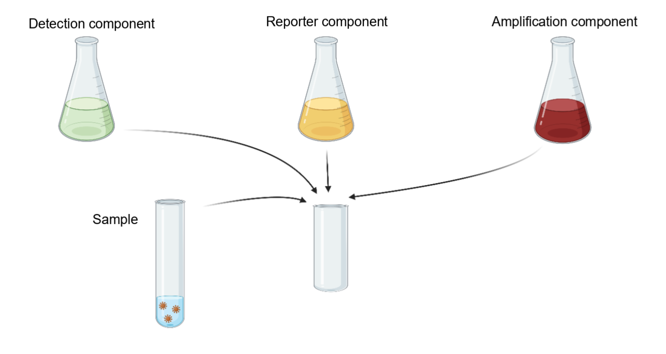
Figure 6: Application of LionDetect – The three cell lysates of the three components are brought together with the sample and enable detection by means of a colour change.
iGEM Team Braunschweig
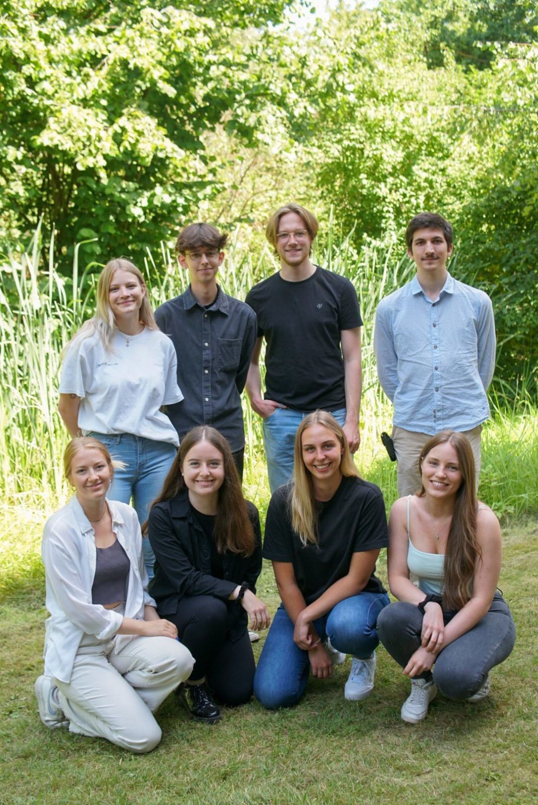
Our team consists of eight students from the Technical University of Braunschweig. We are bachelor and master students in biology and everyone contributes with their skills to the realisation of the project. Within the group, we formed small groups that deal with different aspects of our project: Wet lab, dry lab, sponsoring, social media etc.
If you want to get in contact with us and learn more about our project, visit our website at https://www.tu-braunschweig.de/en/igem/2022 or follow us on Instagram @igem_tu_braunschweig. If you want to know more about our cancer awareness campaign visit us on this Instagram account and learn more: igem_spreads_awareness_

Written by Fabienne Spang, Sina Huxhagen, Melina Nowak, Benjamin Harder and Dr. Andreas Ebertz.