Authenticate the cell line you are working with

Cell lines are essential for research and development! They provide relatively easy to handle, inexpensive and durable systems that mirror in-vivo conditions. From the early 20th century, animal cell lines have been used for studies in medicine, cell biology, genetics, cancer research and drug development. Cell lines that were obtained from major cell culture collections or fellow researchers have since been used for the development of vaccines, drugs, reconstructive medicine and HIV treatment and testing. In 1951, the widely known HeLa cell line was isolated from the cervical cancer biopsy of Henrietta Lacks. This immortalised cell line was the first one to grow exceptionally well in the laboratory. It played a vital role in the development of the first polio vaccine and the discovery of telomerase in human cells. Later, various other cell lines were developed from different tissue sources.

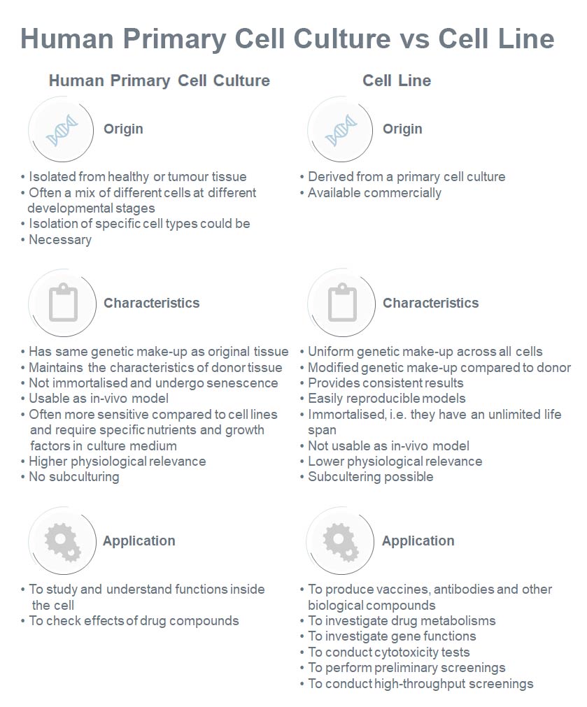
Fig 1: Difference between human primary cell culture and cell line (Oyeleye, O. et al., 2016)

Contamination and misidentification of cell lines

It was during the year 1968 when researchers first identified cross-contamination of cell lines. The researchers noted that overall characteristics and behaviour of their cell lines changed over time Eventually, they determined that contaminating agents such as adventitious microbes or other cell lines caused the changes. It has been estimated that a large proportion of the utilised cell lines are affected by the persistent problem of contamination or are even misidentified. Research results based on contaminated or misidentified cell lines most likely lead to false interpretations, correlations and conclusions (da Silva, 2018; Routray, et al., 2016). Additionally, reported studies carried out with misidentified or contaminated cell lines lead to questionable subsequent studies (Almeida et al., 2016; Horbach and Halfman, 2017).

Here are the facts: About 80,000 cell lines have been developed and, 0.6% (488) of these was reported as misidentified. However, the estimate of misidentified cell lines in use is much higher.  Researchers keep on using misidentified cell lines even after they are unmasked. This has been observed with the human prostatic cancer cell lines JCA-1 and ALVA-31, as well as several thymic cell lines. Already in 1981, a report suggested that a large number of cell lines are contaminated with HeLa (more than 190 entries) followed by T-24 (20 entries) and HT-29 (18 entries) (Horbach and Halffman, 2017). The cell lines HEp-2 and INT-407, for instance, are reported to have been completely replaced by HeLa (Bian et al., 2017; Neimark, 2015). So are you sure that your cell line is what it is supposed to be? Are you confident in the results of your study?

Cell line authentication – Needed for publishing, but how to do it?

Many leading peer reviewed journals such as Nature, AACR Journals, Cell Biochemistry and Biophysics, etc. now require proof of cell line authentication before the submission of cell culture-based research articles. The American Type Culture Collection (ATCC) and American National Standards Institute (ANSI) have documented (ASN-0002) the whys and hows of human cell line identification. The ATCC recommends periodic morphology check via microscope, growth curve analysis, mycoplasma detection and Profiling of Short Tandem Repeats (STR).

When to authenticate your cell line?

In the past, techniques like isoenzyme analysis and karyotyping were used for cell line identification. However, these techniques were not efficient enough to discriminate at the intraspecific level. STR profiling, however, is a reliable, low cost and easy method of cell line authentication. When a lab receives a cell line, relevant data regarding its origin, growth characteristics and culture conditions should be obtained (Reid et al., 2013). Recommended intervals for STR profiling are:

  • When a new cell line is derived from a patient
  • When a cell line is acquired from a non-repository source
  • Before initiating new experiments
  • Before submission of research work for publication
  • When change in morphology and characteristic behaviour of the cell is observed

How to authenticate your cell line

STR profiling establishes a very unique fingerprint of every human cell line (or any other species). STRs (or microsatellites) are about 2-10 bp long and are the hypervariable regions present in mammals, bacteria and fungi as well as in insects, reptiles and fish. STR profiling has the power to discriminate against different individuals of the same species. The numbers of repeats present at the STR loci in each individual varies and give rise to a unique fingerprint. The ANSI/ATCC standard ASN-0002 has mentioned STR profiling as the preferred method for human cell line authentication. A plethora of research work has already been done for the establishment of DNA fingerprinting to carry out the forensic analysis.

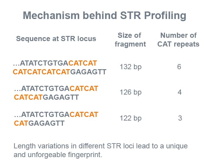
Fig 2: Illustration of the principle behind using STR profiling for DNA fingerprinting

A step by step guide to cell line authentication

The ANSI/ATCC standard ASN-0002 recommends the use of at least eight STR loci (TPOX, TH01, vWA, D16S539, CSF1PO, D7S820, D5S818 and D13S317) and Amelogenin for identification. The process involves the extraction of DNA, amplification via PCR, capillary electrophoresis, conversion of PCR fragments of varying sizes to alleles and comparison to the baseline STR profile of the donor.

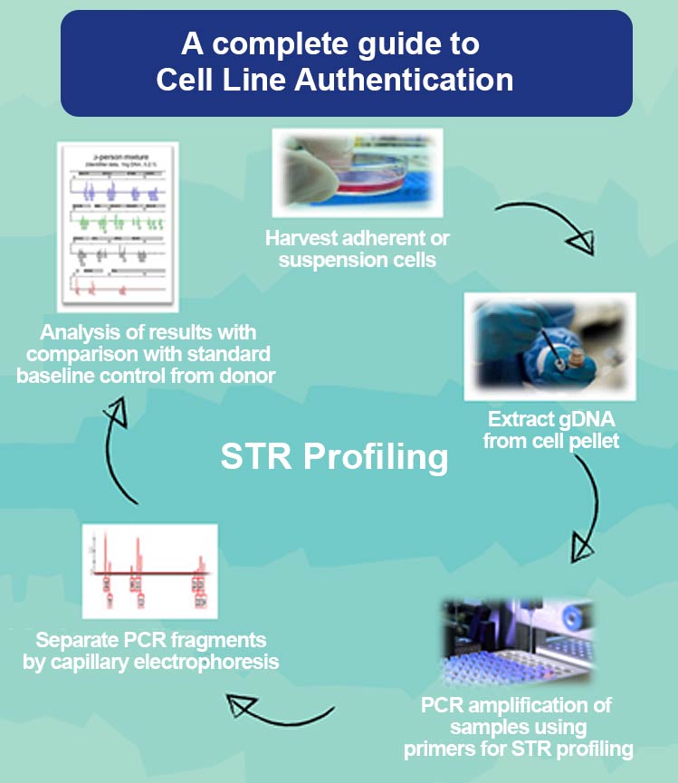
Fig 3: An illustration of the major steps involved in STR profiling.

You can outsource this tedious work of cell line authentication to Eurofins Genomics. We have 20 years of experience in preparing human and animal STR profiles. Our reliable Human Cell Line Authentication service (CLA) is performed following the ISO 17025 standard guidelines and, of course, achieves genotyping according to the ANSI/ATCC standard ASN-0002. The service also includes detection of mycoplasma contamination.

You can now order the CLA as prepaid service online in the Eurofins Genomics web shop. For your convenience, you just need to affix the prepaid barcodes to the reaction tubes and place them in a DropBox. If there is no DropBox nearby, please send your samples by mail. You will receive your high resolution reports in your Eurofins Genomics web shop account ready to download within 5 working days. As work is often very busy and you probably have many things at your mind. In your Eurofins Genomics web shop account, you can set a monthly to yearly reminder option during check out in your Eurofins Genomics web shop account or in “MyPreferences” for cell line authentication. Reminder can also be of a MycoplasmaCheck (MPC).

By Tamseel Fatima and Dr Andreas Ebertz

References
Almeida, J.L., Cole, K.D., Plant, A.L. (2016) Standards for cell line authentication and beyond. PLoS Biol. 14(6): e1002476.
Bian, X., Yang, Z., Feng, H., Sun, H., Liu, Y. (2017) A Combination of Species Identification and STR Profiling Identifies Cross-contaminated Cells from 482 Human Tumor Cell Lines. Sci Rep. 7(1): 9774.
Teixeira da Silva J. A. (2018) Incorrect cell line validation and verification. Teixeira da Silva JA. Incorrect cell line validation and verification. Ann Transl Med. 6(7): 136.
Horbach, S., and Halffman, W. (2017) The ghosts of HeLa: How cell line misidentification contaminates the scientific literature. PloS one 12(10): e0186281
Neimark, J. (2015) Line of attack. Science 347(6225): 938-40.
Oyeleye, O. O., Ogundeji, S. T., Ola, S. I., Omitogun, O. G. (2016) Basics of animal cell culture: Foundation for modern science. Biotechnology and Molecular Biology Reviews 11(2): 6-16.
Reid, Y., Storts, D., Riss, T., Minor, L. (2013) Authentication of Human Cell Lines by STR DNA Profiling Analysis. In: Sittampalam GS, Grossman A, Brimacombe K, et al., editors. Assay Guidance Manual [Internet]. Bethesda (MD): Eli Lilly & Company and the National Center for Advancing Translational Sciences; 2004-. Available from: https://www.ncbi.nlm.nih.gov/books/NBK144066/
Routray, I., Mahmood, A., Ngwa, N. E., Tasleem, M., Sahin, K., & Kucuk, O. (2016) Cell Line Cross-Contamination and Accidental Co-Culture. J Stem Cell Res Ther. 1(5): 00031.

Leave a Reply